Introduction:
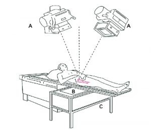
Radiostereophotogrammetric Analysis (RSA) stands at the forefront of modern medical imaging. It provides detailed insights into the dynamics and stability of human tissues and implants. Within this realm, tantalum markers have emerged as invaluable tools. These markers also impact the precision and accuracy of RSA studies.
This article is going to discuss the impact of tantalum markers in RSA studies. Hope that you can have a better understanding.
Understanding RSA Studies:
RSA employs high-resolution X-ray imaging and stereo-photogrammetry. It aims to precisely measure the three-dimensional movement of tissues or implants within the human body. This technique aids in assessing joint replacements, bone fractures, and soft tissue movements with unparalleled accuracy.
Understanding Tantalum Markers:
Tantalum markers play a pivotal role in modern medical processes. These devices offer unparalleled visibility and precision.
They are often crafted from tantalum tubing or tantalum-platinum-iridium capillaries. They serve as radiopaque indicators during intricate catheter-based surgeries. Their distinct visibility under fluoroscopy and imaging technologies allows healthcare professionals to precisely track and navigate catheters. They also ensure accurate placement and navigation within the intricate vascular system.
Advantages of Tantalum Markers:
One of the key advantages of tantalum markers lies in their radiopacity. This feature enables clear visualization under X-ray and fluoroscopic guidance. This heightened visibility is crucial in procedures such as angioplasty, stent placement, and other medical processes.
Moreover, Ta comes with biocompatibility and inert nature. It can work with the human body. This feature minimizes the risk of adverse reactions. Therefore, tantalum markers have become a preferred choice for long-term or temporary implantation.
Besides, they possess precision and safety. Thus, tantalum markers serve as crucial tools in medical procedures.
Applications in Clinical Practice:
Tantalum markers have been applied to RSA and used in many procedures.
1. Orthopedics and Joint Replacements:
In orthopedic surgery, tantalum markers help precise assessment of joint replacements. They are placed on prosthetic components or bones. These markers enable the monitoring of micro-motions and ensure assessment over time.
2. Bone Healing and Fracture Management:
Tantalum markers aid in studying bone healing processes. They are strategically implanted near fracture sites. So, they allow for accurate measurement of healing progression. They also provide valuable insights for treatment evaluation.
3. Soft Tissue Dynamics and Kinematics:
Tracking soft tissue movements is crucial in understanding musculoskeletal function. Ta markers placed on ligaments or soft tissues assist in analyzing joint kinematics. They aid in the diagnosis and treatment planning of injuries or conditions affecting mobility.
4. Research and Development of Implants:
Their use extends to the development of new implants. Tantalum markers integrated into prototype implants help researchers assess performance in simulated conditions, so they can refine designs for improved efficacy before clinical trials.
5. Pediatric Orthopedics and Growth Studies:
In pediatric orthopedics, these markers contribute to understanding skeletal growth patterns. They are placed at specific growth centers. These markers enable longitudinal studies and guide treatment decisions in children with orthopedic conditions.
Challenges and Future Directions:
While tantalum markers have significantly advanced RSA studies, challenges such as marker migration or visibility in certain tissues persist. Ongoing research aims to improve marker design, explore alternative materials, and develop innovative imaging techniques to address these limitations.
Conclusion:
The integration of tantalum markers within RSA studies has reshaped the landscape of human tissue analysis. Such a use offers unprecedented insights into the dynamic behavior of tissues and implants. Their continued evolution and application hold promise for furthering our understanding and enhancing patient care in orthopedics and beyond.
There are a variety of tantalum marker bands available at Stanford Advanced Materials (SAM). They serve as radiopaque markers for various medical procedures and ensure visibility and precision. SAM also offers a range of marker bands made from Platinum, Gold, and Palladium variants. Send us an inquiry if you are interested.
Reference:
[1] Embden, Daphne & Stollenwerck, Guido & Koster, Lennard & Kaptein, Bart & Nelissen, Rob & Schipper, Inger. (2015). The stability of fixation of proximal femoral fractures. The bone & joint journal. 97-B. 391-7. 10.1302/0301-620X.97B3.35077.